An enhancer-dependent chromatin rosette is a specialized, three-dimensional (3D) loop-like DNA structure that physically clusters a gene’s active enhancer with multiple core regulatory elements and its target promoter. This structural framework serves two main purposes: maximizing gene expression and shielding the gene from gene-silencing machinery.
The discovery of the chromatin rosette explained exactly why small CRISPR gene-therapy edits (like those used in the sickle cell disease drug Casgevy) are capable of permanently shutting down a whole target gene.
How the Rosette Functions
- Multi-Way DNA Contact: Instead of a simple single-loop connection between one enhancer and one promoter, the DNA folds into a flower-like “rosette” shape. The enhancer acts as the central hub, drawing in multiple surrounding DNA regulatory segments simultaneously.
- Epigenetic Insulation: The tight rosette fold creates a physical barrier (an “insulated neighborhood”). This structural wall permits high-level transcription and blocks nearby repressive Polycomb proteins from entering and applying silencing epigenetic marks.
- Dependence on eRNAs: The formation and structural integrity of this rosette rely entirely on enhancer RNAs (eRNAs). These are non-coding RNA molecules transcribed directly from the enhancer sequence that act as a structural glue to stabilize the 3D loops.
The Real-World Example: BCL11A and Sickle Cell Disease
The primary model used to identify the rosette is the BCL11A gene. BCL11A is the cellular “switch” that shuts off fetal hemoglobin production and turns on adult hemoglobin.
- In normal blood cells: The BCL11A enhancer generates eRNAs, forming a robust chromatin rosette. This structure keeps BCL11A highly active, which successfully keeps fetal hemoglobin turned off.
- During gene therapy: CRISPR makes a precise cut in the enhancer. This slight sequence disruption stops the production of eRNAs, causing the entire 3D rosette to collapse. Without the protective rosette, repressive proteins rush in, silences BCL11A, and safely flips the switch back on to make therapeutic fetal hemoglobin.
Cutting the BCL11A enhancer using CRISPR therapy disrupts its function by destroying its three-dimensional DNA architecture, which depends heavily on eRNAs.
Here is exactly how this process works at the molecular level:
- The Chromatin Rosette Structure: In healthy adult red blood cell precursors, the intact BCL11A enhancer folds into a complex, multi-loop 3D structure known as an enhancer-dependent chromatin rosette. This structure holds the DNA in a way that provides “epigenetic insulation,” keeping the BCL11A gene highly active.
- The Role of eRNAs: The transcription of this enhancer region produces specific eRNAs. These eRNAs are absolutely essential for the physical integrity of the chromatin rosette because they facilitate the loading of cohesin complexes (like NIPBL-dependent cohesin) that lock the 3D loops in place.
- The CRISPR Cut: When CRISPR-Cas9 cuts this enhancer by targeting GATA1 binding sites (second diagram below), it alters the DNA sequence and prevents it from properly transcribing the eRNAs (first diagram below).
- The Downstream Effect: Without the eRNAs, cohesin can no longer properly bind to the region. This causes the rosette structure to collapse. Once the protective 3D structure unfolds, the BCL11A gene is exposed to repressive proteins that silence its expression.
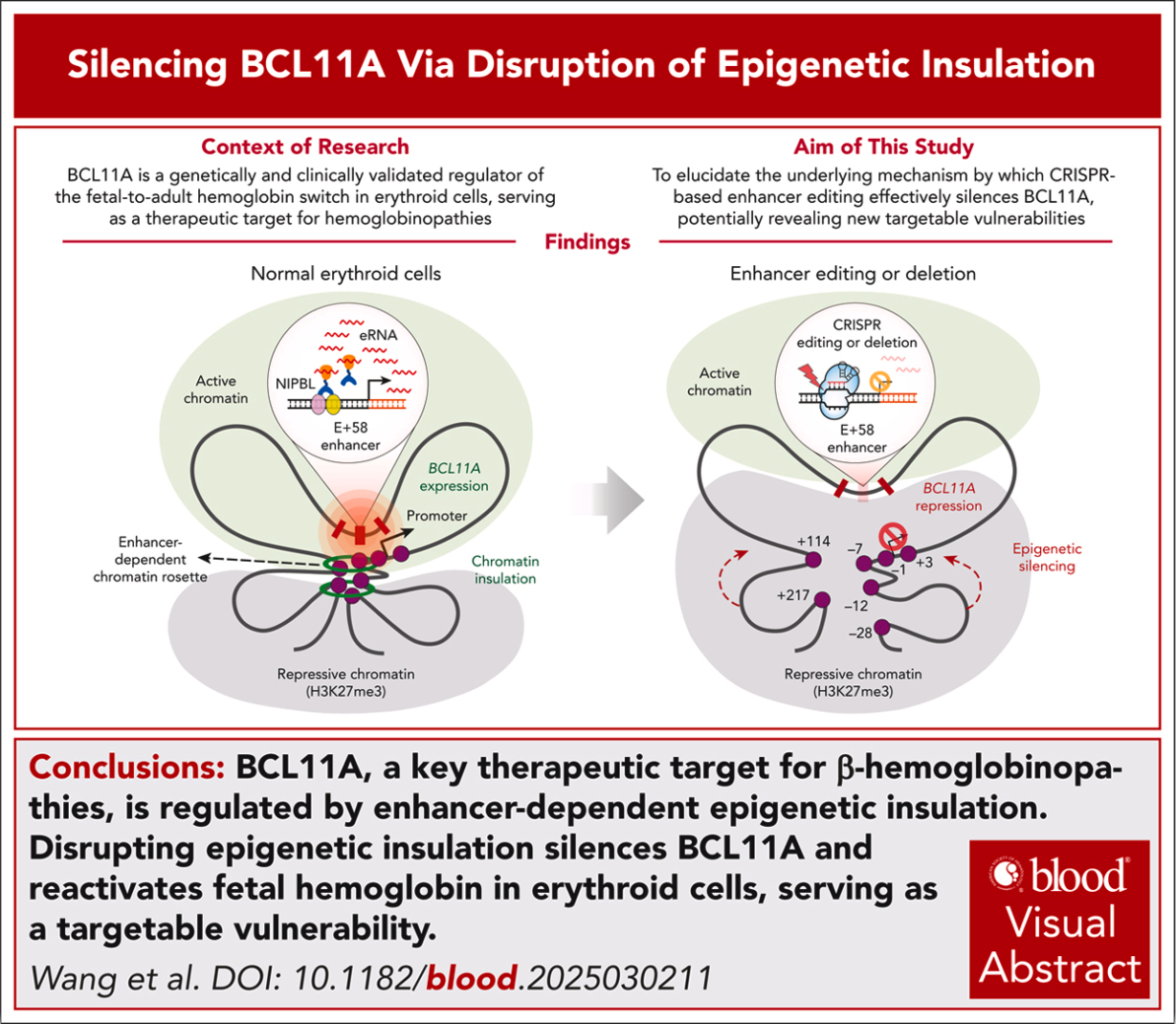
Reference: Wang K. et al. Silencing of BCL11A by disrupting enhancer-dependent epigenetic insulation. Blood (2026) 147 (13): 1470–1484.
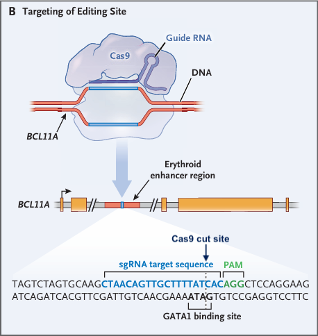
Reference: Frangoul H. et al. CRISPR-Cas9 Gene Editing for Sickle Cell Disease and Beta-Thalassemia. N Engl J Med 2021; 384:252-260.
Because BCL11A is a master repressor of fetal hemoglobin, disabling this enhancer means the gamma-globin genes are no longer silenced, ultimately producing therapeutic levels of fetal hemoglobin (HbF) to treat sickle cell disease and beta-thalassemia.
To summarize:
1. The Chromatin Rosette Structure
In healthy red blood cell precursors, the BCL11A enhancer (like the +58kb region) loops onto itself and nearby elements to form a 3D enhancer-dependent chromatin rosette. This highly insulated conformation keeps the BCL11A gene accessible and highly active.
2. The Role of eRNAs
Transcription of the enhancer region produces enhancer RNAs (eRNAs). Recent biological studies have proven that these eRNAs are critical structural molecules; they facilitate the loading of cohesin complexes (like NIPBL-dependent cohesin) which literally lock the complex 3D chromatin loops in place.
3. The CRISPR Cut
When CRISPR-Cas9 is used to target and disrupt the BCL11A enhancer, it fundamentally alters the DNA sequence. This mutation damages the transcription machinery, preventing the enhancer from properly producing its vital eRNAs.
4. The Downstream Effect
Without these eRNAs, cohesin can no longer bind efficiently to the enhancer region. The “rosette” structure subsequently collapses, causing the BCL11A gene to lose its protective “insulation”. Once exposed, repressive proteins enter and permanently silence the BCL11A gene.
5. Clinical Result
BCL11A normally acts as a master silencer of fetal hemoglobin in adults. By disabling the enhancer, BCL11A is turned off, allowing the gamma-globin genes to remain active. This produces therapeutic levels of fetal hemoglobin (HbF), which compensates for the mutated adult hemoglobin, preventing red blood cells from sickling.
This exact biological mechanism is the foundation of FDA-approved gene therapies like Casgevy (developed by CRISPR Therapeutics and Vertex Pharmaceuticals).